Harnessing Science to Advance Human Health

Innovative research in immunology, epigenetics and genomics will help realize the promise of precision medicine.

Illustrations by Wenting Li
February 14, 2022 By Mark Witten
View PDF version

Advances in improving human health often arise out of the discoveries made by talented researchers working in the fundamental sciences.

Translational breakthroughs in applying these findings to develop new and more effective medical treatments are made possible through the ingenuity of basic scientists, who invent better tools and techniques to move their research forward in diverse fields such as immunology, epigenetic regulation, functional genomics and oncology.

The leading-edge life sciences research of three former Azrieli Fellows—Deborah Winter at Northwestern University in Chicago, Ziv Shulman at the Weizmann Institute of Science, and Oren Ram at the Hebrew University of Jerusalem—shows the promise their innovative work holds for improving human health.

Share this article

Each scientist is helping to lay the foundations for precision medicine in different ways. Through advances such as Winter’s genomic profiling of macrophages in patients with autoimmune disease, Ram’s epigenetic profiling of rare treatment-resistant cancer cells, and Shulman’s whole-organ imaging of the gut immune system, these early-career scientists are opening new avenues for personalized, targeted treatments to improve outcomes in diseases such as cancer, rheumatoid arthritis, liver disease, bacterial enteric infections and coronavirus infections.

Deborah Winter, Comutational Immunologist
Principal investigator, Winter Lab of Functional Genomics
Northwestern University

Computational immunologist Deborah Winter, left, with some current and former members of her research team at the Feinberg School of Medicine at Northwestern University in Chicago.

To do precision medicine and develop targeted treatments, you need to first understand how the genomic landscape is altered for the disease in your therapeutic sights.

When Deborah Winter began her postdoctoral research at the Weizmann Institute of Science in Israel in 2013, the universe of macrophages—cells that detect and destroy pathogens—in the body’s immune system at the genomic level was relatively unmapped and poorly understood. Scientists in immunology didn’t know how much variation there was in gene expression patterns for macrophages in different tissues, or how these vital pathogen-gobbling cells develop tissue-specific functions, such as lung macrophages metabolizing lipids and brain macrophages pruning synapses in neurodevelopment.

As an Azrieli International Postdoctoral Fellow in Ido Amit’s immunogenomics lab at the Weizmann Institute from 2013 to 2016, Winter was encouraged to be bold and fully apply her computational biology skills, as well as her expertise in epigenomics and functional genomics, to answer important questions in immunology, a field that was new to her. The work involved using mouse models to examine how macrophages in different tissues vary in how they organize DNA into chromatin; this, in turn, affects their function.

“It was important to understand how genes were regulated in macrophages in each tissue, and how macrophages in the lungs, heart or brain know what they are supposed to do. No one knew then how macrophages function at the genomic level when healthy, and what happens when something goes wrong,” says Winter, who in 2016 became an assistant professor in the Division of Rheumatology at Northwestern University’s Feinberg School of Medicine. “Today, we know that in a lot of inflammatory and autoimmune diseases, new populations of harmful macrophages arise that we can potentially target.”

Winter is the co-first author of a landmark study on macrophages that was published in Cell in 2014: “Tissue-Resident Macrophage Enhancer Landscapes Are Shaped by the Local Microenvironment.”

Her highly cited paper mapped in comprehensive detail how macrophages develop distinct tissue-specific functions in seven macrophage populations: brain, spleen, liver, lung, peritoneal cavity, large intestine and small intestine.

“We found that the tremendous heterogeneity in gene expression was due to the fact that each macrophage population has a different chromatin landscape. We showed the importance of the local microenvironments and how each macrophage population knows what it is supposed to do. Our study also revealed that tissue-resident macrophages can adopt tissue-specific functions by reprogramming their chromatin landscape in response to signals from the local environment,” Winter explains. “The paper made such a splash, and it was the foundation for work I’ve been doing since on genomic profiling of macrophages and targeting treatments for inflammatory and autoimmune diseases, lung and liver disease, and lung infections like COVID-19.”

The specialized training Winter received as an undergraduate in the University of Toronto’s inaugural Bioinformatics and Computational Biology Program from 2004 to 2008 was a springboard to more advanced training as a computational biologist at Duke University in North Carolina. Her research there focused on gene regulation, using high-throughput sequencing assays to study chromatin dynamics across diverse human cell lines. She also worked on the Encyclopedia of DNA Elements (ENCODE), the second-generation human genome project that aims to identify all functional elements in the human genome.

“Genes account for only up to two per cent of the human genome, and we realized that a big function of the other 98 per cent of what used to be called ‘junk DNA’ is gene regulation,” Winter says. “It was a great experience working on basic gene regulation, but for my postdoc, I wanted to get closer to cell biology.”

At the Weizmann Institute, Winter applied computational modelling and single-cell genomic approaches to investigate regulation and gene expression patterns of macrophages in different tissues. While there, she co-authored another highly cited paper that was published in Science in 2016: “Microglia development follows a stepwise program to regulate brain homeostasis.”

Key Terms

Antibody affinity maturation is an evolutionary selection process in which B cells stimulated by helper T cells mature into antibodies with an increased capacity to destroy a particular pathogen. This process is influenced by the affinity, or strength of attraction, between an antibody and a pathogen.

Chromatin is a type of genetic material within chromosomes. It is composed of DNA and proteins. The major proteins in chromatin are histones, which package long DNA molecules into more compact, denser structures.

Epigenetics is the study of how behaviour and environment influence gene activity and expression. Epigenetic changes are reversible and do not alter one’s DNA sequences.

Intravital two-photon microscopy is a fluorescence imaging technique that allows the visualization of biological processes in live animals at depths unachievable with conventional fluorescence or confocal microscopy.

Macrophages are white blood cells in the immune system that engulf and digest cellular debris, foreign substances, microbes and cancer cells in a process called phagocytosis.

Microfluidics involves the study, design and use of devices that can manipulate tiny amounts of liquid to perform various scientific processes. It is increasingly used in the life sciences because it allows researchers to conduct controlled experiments relatively quickly and inexpensively.

Single-cell sequencing allows researchers to study DNA, RNA or epigenetic information from individual cells using optimized next-generation sequencing technologies.

“They don’t kowtow to dogma. My experience at Weizmann was a defining one and made me a bolder scientist.”
Deborah Winter

The research uncovered a three-stage development process for microglia (brain macrophages) to regulate brain homeostasis and showed how disruptions in these pathways may be linked to several neurodevelopmental disorders.

“The Azrieli Postdoctoral Fellowship at Weizmann gave me opportunities to be exposed to different approaches and different types of research, which broadened my horizons. Labs in Israel do very bold science and don’t shy away from controversy and expressing opinions. They don’t kowtow to dogma. My experience at Weizmann was a defining one and made me a bolder scientist,” Winter says.

At the Feinberg School of Medicine at Northwestern, Winter has been applying these computational modelling and single-cell genomic techniques to macrophage samples from human patients to study how genomic variability leads to disease. She focuses on autoimmune diseases, such as rheumatoid arthritis (RA) and scleroderma, an area that urgently needs more tailored and targeted treatments. In RA, macrophages are overactive and produce toxic, inflammatory proteins that destroy joint tissues. Current treatments are trial and error, and are effective for some patients but ineffective for many others who go through 12 weeks of therapy, experience no improvement, and then try another.

“There are various treatments for rheumatoid arthritis, but only about a third of patients respond to the first treatment given, and the disease can progress rapidly in those who don’t respond. We waste about $2.5 billion a year on ineffective therapy for this disease in the United States,” she says.

In early 2018, Winter was co-senior author of “Transcriptional Profiling of Synovial Macrophages Using Minimally Invasive Ultrasound-Guided Synovial Biopsies in Rheumatoid Arthritis” (Arthritis & Rheumatology), a groundbreaking pilot study that could bring precision medicine to the treatment of RA. Using ultrasound-guided biopsies of synovial tissues (lining of the joints) obtained from patients at six U.S. medical centres, Winter and her collaborators were able to characterize patients based on the gene expression profiles of their macrophages. This genomic profiling revealed the stage of disease, which patients had the most severe disease, what biologic therapies patients were on.

Winter’s successful transcriptional profiling of synovial macrophages in RA patients led to a new and larger clinical study, which has enrolled about 100 patients and will include several hundred more. Scientists conduct a biopsy to remove joint tissue from patients at the start of a new therapy, and they do another biopsy six weeks later to see if they can find a predictive signature of gene expression that clearly identifies which patients respond to a particular therapy.

“What we’re doing is translational. Our goal is to come up with macrophage biomarker profiles to predict which patients will respond to a particular treatment. You need to appreciate how patients vary from each other to be successful at precision medicine, which is evidence-based and personalized,” she says.

Winter is now applying these functional genomic approaches to develop precision medicine treatments for pediatric liver disease and scleroderma as well. In a co-authored study, “Transcriptional profiling of pediatric cholestatic livers identifies three distinct macrophage populations” (PLOS ONE, January 2021)she and her colleagues identified three distinct macrophage populations in tissue samples from patients with pediatric cholestatic liver disease (blocked or reduced bile flow). “These findings may allow for future development of targeted strategies to reprogram macrophages and promote a population of good macrophages to work as a treatment,” she says.

Currently, the Winter Lab is preparing to publish a study focusing on scleroderma, which was initially described in a 2019 abstract: “A Common Transcriptional Signature Is Present in Circulating Classical Monocytes and Skin Macrophages in Systemic Sclerosis.” Scleroderma is a disease that affects skin as well as multiple internal organs that are hard to biopsy, including the lungs, heart and kidneys. An alternative approach using patient blood samples to inform clinical decision-making would be invaluable for detecting characteristics of systemic disease, such as which organs were involved, speed of progression and likelihood of treatment response. Winter identified a gene signature in circulating blood monocytes (macrophage precursors) that could predict the progression of scleroderma in a patient and help guide treatment. “You could come up with a personalized treatment plan, based on differences in the gene expression signature of circulating monocytes, which would allow you to monitor how quickly the disease is progressing and the need for aggressive intervention,” she says.

“I love what I do. I discovered in my postdoc, with my first exposure to immunology, that I like learning on the job about a new field from scratch.”
Deborah Winter

Winter’s experience doing postdoctoral research at the Weizmann Institute was a catalyst for applying her innovative methods more widely to human health and targeting treatments for patient subpopulations in many diseases. “I love what I do. I discovered in my postdoc, with my first exposure to immunology, that I like learning on the job about a new field from scratch. I knew little about human disease and rheumatoid arthritis at first, but I learned on the job,” she says. “I want to collaborate with people who have expertise in each disease. I have a unique perspective, which I can bring into new fields and have an impact.”

Ziv Shulman
Principal Investigator, The Shulman Lab
Weizmann Institute of Science

Ziv Shulman’s novel technique for visualizing the whole organs of the human immune system illuminates more clearly how protective antibodies are generated to protect against pathogens. Photograph by Shauli Lendner
“Imaging through live microscopy is very exciting and stimulating because you get to watch the antibody immune response and dynamics with your own eyes...”
Ziv Shulman

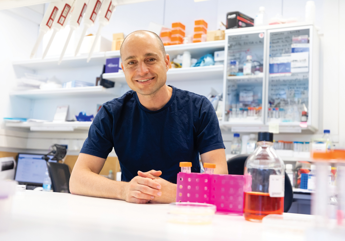
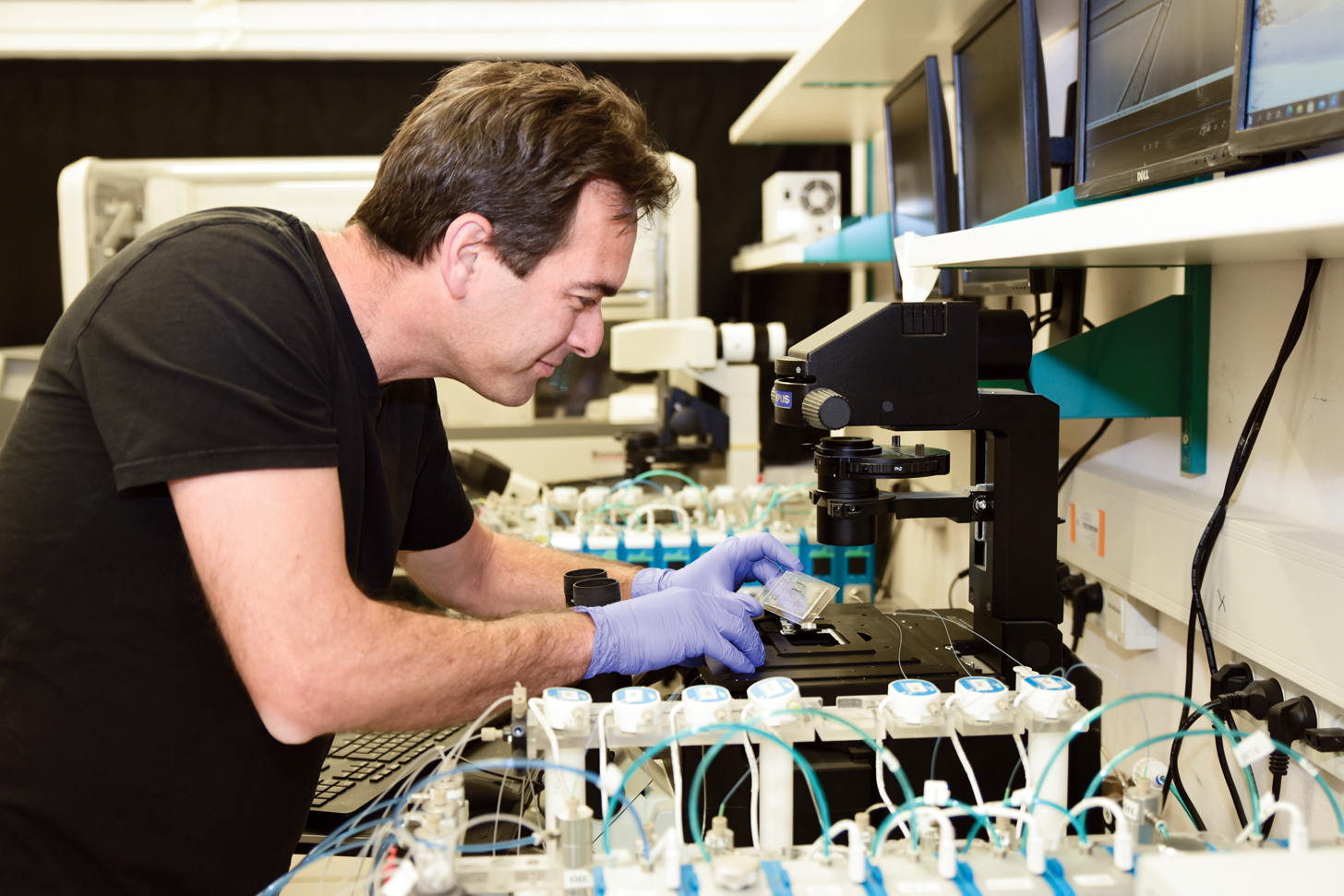
Watching the immune system in action has been key to Ziv Shulman’s discoveries about how the body’s peripheral lymph system and gut immune system make antibodies precisely targeted to fight pathogens in very distinct ways. Shulman has invented and applied cutting-edge visualization tools and techniques to show how the evolutionary selection process generates the most protective antibodies against a particular pathogen.

“Imaging through live microscopy is very exciting and stimulating because you get to watch the antibody immune response and dynamics with your own eyes,” says Shulman, principal investigator and head of The Shulman Lab, which focuses on cellular dynamics and molecular regulation of the adaptive immune response.

As an Azrieli Faculty Fellow in the Weizmann Institute’s Department of Immunology from 2016 to 2020, Shulman saw an urgent need for a better way to visualize the poorly understood gut immune system. The specialized immune niches, or sites, within the system’s lymphoid organs are so small and well hidden that scientists have found it difficult to study them with standard imaging methods.

“I thought, if you can image a whole brain in neuroscience, why can’t you image the whole organs of the immune system? We learned how to make the gut lymphoid organs transparent using light-sheet fluorescence microscopy and capture all of them to visualize the gut immune system in a more holistic way at single-cell resolution,” Shulman says. “In one shot, you see everything, and we were able to capture small immune niches that are difficult to detect using conventional microscopy. That was a big advance, which allowed us to ask important questions about how the immune response in the gut works.”

In the breakthrough paper “BCR affinity differentially regulates colonization of the subepithelial dome and infiltration into germinal centers within Peyer’s patches” (Nature Immunology, March 2019), Shulman and his colleagues described using whole-organ imaging to show how the gut immune system’s B cells—a type of white blood cell—play by a different set of rules than those in the peripheral lymph system. They visualized the gut lymphoid organs of mice that had been immunized orally to reveal the gut immune response in action. While the peripheral immune system aims to select and produce the most effective, high-affinity antibodies quickly, the gut system antibody response involves a slower two-stage process.

“In the first stage, affinity-based selection is delayed, and both low- and high-affinity antibodies are produced. For the second stage, the immune cells must migrate into niches in which the proper antigens have accumulated over time, so that affinity training can take place to produce high-affinity antibodies,” Shulman says. “But unlike in the peripheral lymph system, the antigen levels within the gut immune system’s niches are mostly too low to stimulate efficient antibody generation.” He discovered that an area of the gut’s lymphoid organs, known as the subepithelial dome, plays a crucial role in their antibody immune response. These findings could help scientists design new and better oral vaccines.

“Our study suggests that an antigen needs to be targeted to the subepithelial dome to trigger an effective immune response. Good targeting will increase the dose of the vaccine and promote a more effective immune response in the gut. Better targeting can be used in vaccinations against pathogens that enter through mucosal tissues, such as rotavirus, HIV, polio, and bacteria like salmonella,” he says.

Shulman’s interest in immunology research was sparked in 1999 after he persuaded an Israeli biotech company to hire him as a technician. “I got to hang around the lab and see what they were doing. Their goal was to cure spinal cord injuries using the body’s immune system. It was fascinating, and I wanted to be like that,” Shulman says. The experience inspired him to start a bachelor’s degree in animal science at the Hebrew University of Jerusalem in 2001. His subsequent cutting-edge research on immune cell migration and adhesion at the Weizmann Institute earned him prizes from the Feinberg Graduate School for outstanding achievements, once as an MSc student in 2006 and again as a PhD student in 2011.

While completing his PhD research, Shulman used live imaging microscopy to learn about how immune cells pass through cell layers lining blood vessels. Shulman’s and the lab team’s findings were published in the article “Transendothelial migration of lymphocytes mediated by intraendothelial vesicle stores rather than by extracellular chemokine depots” (Nature Immunology, December 2011). “I could visualize the immune cells’ tiny, sticky legs moving like a millipede. Live imaging was key to the discovery, and you can’t see it by using traditional microscopy that produces static pictures,” explains Shulman, who did postdoctoral research at Rockefeller University in New York City to learn more advanced imaging techniques and observe how high-affinity antibodies are formed.

Forming efficient antibodies against specific pathogens involves a biological selection process called antibody affinity maturation, in which B cell antibody genes randomly mutate. During his postdoc, Shulman developed a novel system to visualize and analyze how T cells and B cells interact in real time to form the best antibodies against future pathogens in response to vaccination. He used intravital two-photon laser scanning microscopy and automated quantification algorithms to shed light on the process.

In papers published in Science in 2013 (“T Follicular Helper Cell Dynamics in Germinal Centers”) and 2014 (“Dynamic signaling by T follicular helper cells during germinal center B cell selection”), Shulman described the intricate molecular dance between T and B cells after infection, and he showed how these two types of immune cells from numerous short-term contacts to prepare the antibodies and establish long-lasting protection. “You activate the microscope and it’s like watching a show on stage. We saw the dynamics of many short-term interactions that happen over hours, with B cells being directed by T cells, and T cells learning from their interactions with B cells,” explains Shulman, whose work earned him Rockefeller University’s Tri-Institutional Breakout Prize for Junior Investigators in 2015.

In addition to his current gut immunity research pointing to new ways of developing oral vaccines, Shulman is working with recovered COVID-19 and cancer patients to develop therapeutic antibodies for these conditions. His team has developed a screening strategy to detect both anti-corona antibodies in COVID-19 patients and autoantibodies in the ascites—abnormal buildup of fluid in the abdomen—of patients with ovarian or pancreatic cancer. This strategy aims to generate anti-corona and antitumour antibodies for new patient treatments and for use with existing immunotherapy approaches.

Oren Ram, Epigenomicist
Principal Investigator, Epigenomics Ram Lab
Hebrew University of Jerusalem

Epigenomics researcher Oren Ram has developed an innovative singlecell sequencing technology to better detect and analyze treatment-resistant cancer cells. Photograph by Boaz Perlstein

Drug resistance remains one of the biggest challenges in cancer therapy. A patient with advanced cancer is given a treatment that helps shrink their tumour, but then weeks or months later the cancer comes back, and the drug no longer works. Research has shown that cancers often become resistant to therapy due to both genetic and epigenetic differences in the small subsets of tumour cells left behind after treatment.

To learn more about these drug-resistant subsets, Oren Ram, an Azrieli Faculty Fellow at the Hebrew University of Jerusalem and head of the school’s Epigenomics Ram Lab, has developed innovative single-cell sequencing tools using drop-based microfluidics. The tools can detect and characterize rare subsets of cancer cells in tumours that resist drug treatment and drive the disease’s progression.

While existing single-cell sequencing technologies can reveal the scope of heterogeneity in cell populations, they lack the sensitivity to fully characterize aspects of cell heterogeneity and detect subtle but important epigenetic states in these rare cancer cells. “We sequence in bulk to identify mutations and aberrations in cancer cells that drive the cancer. But these rare mutations are difficult to spot, and if you only look at a single cell, you don’t have enough information about the cancer cell’s genetic mutation or its epigenetic state,” says Ram, whose lab is in the Department of Biological Chemistry at the Hebrew University’s Alexander Silberman Institute of Life Science.

To overcome the limitations of single-cell-based assays, Ram developed a novel technology that uses droplet-based RNA sequencing to grow single cells into small clones. “With our CloneSeq platform, a rare mutation is no longer rare because we can grow single cells in 3D hydrogels and produce up to 50 cells in a clone. This amplifies the signal and gives us more information about the cell’s mutation, gene expression level and replication rate,” he says.

Ram explains the platform’s unique capacity to discover and profile rare and previously hidden subpopulations of cancer cells in a co-authored paper currently under revision by Developmental Cell: “CloneSeq: A Highly Sensitive Single-cell Analysis Platform for Comprehensive Characterization of Cells from 3D Culture.” It describes how CloneSeq analysis of non-small-cell carcinoma cells can detect novel cancer-specific subpopulations, as well as subtle differences in their expression states that can’t be detected with other methods, including cancer stem-like cells, high and low replicative cancer cellular states, and different levels of invasiveness.

Now, Ram is optimizing the CloneSeq technology to profile cells derived directly from cancer patients and help clinicians conduct drug screens for early detection of treatment-resistant cells. He is collaborating with oncologists at Hadassah Hospital – Ein Kerem in Jerusalem to analyze and profile tumours from patients with ovarian cancer and glioblastoma, two of the deadliest and most treatment-resistant cancers.

“In moving towards personalized medicine, we would take biopsied tissue from cancer patients and use Clone RNA-seq assays to get useful information about the genetic mutation and the functional epigenetic state of the treatment-resistant cells,” he says. “This could allow oncologists to test and perhaps add a suitable drug to the treatment regimen that would be targeted specifically to avoid resistance, based on the genetic and epigenetic cellular information.”

Ram was attracted to the exploding field of epigenetics while finishing his PhD in molecular genetics at Tel Aviv University in 2009. In his postdoctoral research at Massachusetts General Hospital, Harvard Medical School and the Broad Institute of MIT and Harvard, Ram focused on the regulation of chromatin, the complex of non-genetic material associated with DNA that drives gene expression. He devised a novel technique, called ChIP-string, as a screen to study and measure the activity of chromatin regulators in different cell types. In “Combinatorial Patterning of Chromatin Regulators Uncovered by Genome-wide Location Analysis in Human Cells” (Cell, December 2011), Ram and the research team revealed specific combinations of chromatin regulator proteins controlling essential chromatin activities, such as histone modification.

“The ChIP-string technique allowed us to learn how 30 chromatin regulators work in two human cell types—cancer cell lines and embryonic stem cells. It was fascinating to better understand how the chromatin regulation underlying all cell types works,” Ram says.

“We want to better understand gene regulation dysfunction during cancer development and aging.”
Oren Ram

His next challenge was to develop a method of studying chromatin regulation that would be sensitive to cell-to-cell variation. Inspiration struck while Ram was playing basketball at Harvard with a physicist, Assaf Rotem, who specialized in microfluidics. After the game, they started talking about the idea of encapsulating single cells in microfluidic drops to allow for single-cell analysis of chromatin states. Together, Ram and Rotem developed a new technique called Drop-ChIP, which combines microfluidics, DNA barcoding and next-generation sequencing to identify distinct epigenetic states within a single-cell population. Ram says Drop-ChIP is an important advance that could provide new insights into the role of cellular epigenetic heterogeneity in both basic biology and disease states such as cancer. The findings were published in “Single-cell ChIP-seq reveals cell subpopulations defined by chromatin state” (Nature Biotechnology, October 2015).

“I got a position at Hebrew University because of that study, since I was bringing a valuable technology to Israel that didn’t exist before,” he says.

As a new faculty investigator, Ram was able to quickly recruit talented students for his lab team and secure the equipment and supplies needed to move his single-cell ChIP-sequencing methods (including CloneSeq) and broader epigenomics research forward. “The Azrieli Fellowship gave me a jump-start to advance the technology and my research more rapidly,” he says.

In 2017, Ram won a five-year European Research Council Starting Grant worth €1.5 million for his research project “Decoding the Epigenomic Regulatory Code by the Use of Single Cell Technologies.” The aim is to further develop his innovative drop-based single-cell microfluidics technologies and apply them to questions about cellular heterogeneity and epigenomic regulation during early differentiation of embryonic stem cells.

In a paper currently under revision for publication in PLOS Genetics, “DNA Methylation Patterns Expose Variations in Enhancer-Chromatin Modifications during Embryonic Stem Cell Differentiation,” Ram and the research team combined microfluidics, cutting-edge ChIP-sequencing and single-cell RNA-sequencing methods to provide new insights into the functional relevance of DNA methylation in the context of enhancers during embryonic stem cell differentiation. Enhancers are regulatory regions of DNA responsible mainly for increasing the possibility of transcription of a certain gene.

“DNA methylation is usually associated with suppression, and enhancers associated with activation. We wanted to better understand the crosstalk between DNA methylation and enhancers, and found that differences in enhancer-specific methylation are associated with and can be explained by cell-to-cell variation,” Ram says. “This gives us a better understanding of dynamic enhancer regulation, which could be useful for investigating dysfunction that occurs in gene regulation during cancer development and aging.”

Innovations in tools and techniques are helping to drive Ram’s research forward in epigenomics, and that is opening doors to potential health applications with collaborators in oncology. “We’re asking unique questions in the basic science of stem cell differentiation, for example, that are difficult to answer unless you have access to these cutting-edge technologies,” he says. “It’s also exciting to work with other medical experts who can utilize these techniques to explore new treatment options for specific cancers that are among the most difficult to treat effectively.”

Have information and updates about the Azrieli Fellows Program sent directly to your inbox.

Subscribe to our newsletter

* indicates required

All fields are mandatory.

Top