Protected: Where Memories Live

There is no excerpt because this is a protected post.

Photographs by Avital Hirsch
October 14, 2026 By Anne Casselman
View PDF version

At the scale of half a centimetre, a mouse’s brain activity resembles a thunderstorm seen from low orbit, a constellation of flashes tracing illuminating patterns through the clouds of brain tissue. We know this because neuroscientist Arseny Finkelstein and his colleagues have offered us a glimpse into the brain using microscopes and neural imaging techniques that capture the activity of tens of thousands of neurons in the mouse brain. “In effect, we built windows into the brain that allowed us to visualize its activity at the resolution of individual cells,” Finkelstein says.

Finkelstein is an assistant professor in the Department of Neuroscience and Brain Disorders at Tel Aviv University and was an Azrieli Early Career Faculty Fellow in the 2021–2022 cohort. Neuroscientists like him devise ingenious techniques to discover how the brain’s network of cells makes decisions and forms memories. Thanks to these efforts, we can now watch enormous networks of neurons in mice as the animals search to locate rewards — enabling the mapping of connectivity during learning.

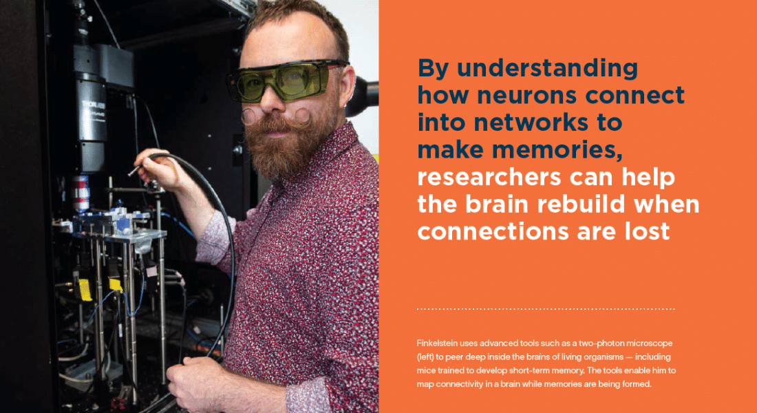
Humble though it may seem, studying these basic building blocks of brain connectivity in mice has enormous implications for our understanding of memory, identity and cognitive decline. With a better understanding of how neurons connect into networks to make memories, researchers can help the brain rebuild when those connections are lost.

Finkelstein’s fascination with memory dates to his childhood, when he revelled in the family’s history recounted by his grandparents. But it wasn’t only the memories and their contents that captivated him so much as the realization that those memories were encoded and retained physically for decades inside the human brain. “Essentially, we have this simulator in our mind that we can always return to and experience many alternative versions of our lives, both in the past and in the future,” he says.

His journey into brain research began at the Weizmann Institute of Science, where he worked on his master’s degree, studying immune-based approaches to stroke and neurodegenerative disease. For his PhD, also at the Weizmann Institute, he joined Nachum Ulanovsky’s lab and shifted into systems neuroscience. There he uncovered how bats understand space in three dimensions. He then spent five years studying decision making as a postdoctoral fellow at Janelia Research Campus, part of the Howard Hughes Medical Institute in the U.S. In 2022, he joined Tel Aviv University, where he established his own lab.

Finkelstein credits the Azrieli Fellowship with crucial support at a pivotal point in his scholarly career. “The Azrieli Fellowship was instrumental in supporting the early days of my lab. It enabled me to concentrate on developing and expanding our approach to mapping memory formation and network changes during learning,” he says. “Given the highly interdisciplinary nature of neuroscience and memory research, interactions with fellow Azrieli scholars were especially enriching and led to valuable exchanges of ideas.”
Share this article

From the beginning, Finkelstein was animated by the idea that our brains can experience a space that’s vast and rich through memory and imagination. Fortunately for him, he and his colleagues can now chip away at answering these foundational questions about what memory precisely is at the neural level, thanks to recent advances in neuroscience. “We can, for the first time, dare to start asking these questions on a mechanistic level,” he says.

In November 2025, Finkelstein and his colleagues published research in the journal Nature that tracked the activity of 100,000 neurons as a mouse licked up sugar water from different locations around its mouth. They were able to document in detail not only the brain activity of 1 square millimetre of the mouse’s motor cortex — part of the cortex, the brain’s outer layer responsible for decision making — but they were also able to study how hundreds of thousands of neurons were linked through more than 20 million pairs of potential connections. It’s a more comprehensive picture of real-time brain activity than ever captured before — and the first map of part of the cortex in a living animal.

Perhaps the study’s most striking finding was the existence of rare hub neurons. While most neurons showed relatively few connections, a small number of highly connected neurons influenced many neighbouring cells — even though hub neurons were not strongly tuned to any particular target location themselves. Finkelstein’s work showed that neurons encoding the same behavioural goal are wired together in a spatial pattern, which may help animals rapidly learn goal-directed actions.

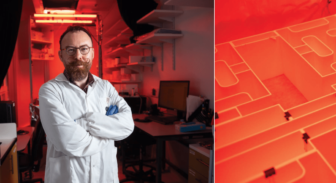
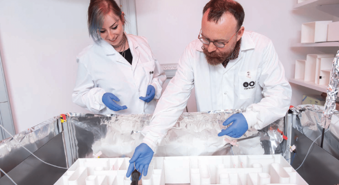
Finkelstein and Adva Danielly, a graduate student in neuroscience, prepare a mouse to navigate a complex maze and pursue goals based on spatial memory. Finkelstein and colleagues have devised ingenious techniques to discover how the brain’s network of cells makes decisions and forms memories. As a result, they can now watch enormous networks of neurons in mice as they locate rewards like sweetened water around their mouths.

Karel Svoboda, now at the Allen Institute in Seattle, was Finkelstein’s postdoctoral advisor at Janelia Research Campus, where Finkelstein conducted the work that led to the Nature paper. “[Arseny] was really pushing this link of looking at neural activity — the symphony of electrical signals in the brain that generate behaviour — and then how that actually relates to connectivity between neurons in the same tissue,” says Svoboda. “That’s unprecedented.”

The end result is much more than an isolated finding. It’s a method by which Finkelstein and his team can work toward his larger mission piece by piece: to understand, for the first time, the physical embodiment of memory inside the brain. Neuroscientists know that memory involves changes to the connectivity between neurons, but any more detail than that — such as which neurons hold memory through these connections or how connections change as new memories form — and the picture gets murky.

The first step to answering this question involved mapping the mouse’s brain at one time point, a task that Finkelstein has now completed. Next, he and his team are applying those same methods to map neuronal connections in one mouse’s brain at different time points during learning to capture the changes that learning precipitates in the mouse’s connectome, essentially the brain’s wiring diagram.

Finkelstein’s research showed that neurons that represent the same sugar water location relative to a mouse’s snout tend to form strong local connections. But when those neurons are farther apart, they are more likely to inhibit one another. He says this pattern of connections could be the physical basis for how the brain briefly holds onto the memory of where something is located.

Finkelstein and his team are now testing the idea in the lab. This involves teaching mice to use their short-term memory to make decisions and then tracking any resulting changes to an individual mouse’s neural network. As they teach the mouse and increase its capacity for short-term memory, they simultaneously track any changes to its cortical neural network over time.

“This is an exciting moment for neuroscience, but we are still in front of a big, giant mystery.”

“Understanding how these processes are implemented in the brain is very important to understand what happens to the network when it’s breaking down, when memories are breaking down, when we’re left in this sort of mental palace with bare walls,” Finkelstein notes.

While the idea of training short-term memory in a mouse might seem a far cry from doing so with an Alzheimer’s or stroke patient, understanding how neural networks form short-term memory is a critical step in understanding how to stabilize or salvage a compromised neural network in humans. Following a stroke, part of the brain’s network dies abruptly, and the remaining parts of the brain’s network must learn and reassign roles to compensate for the loss.

Finkelstein is using the same approach of connectivity mapping to understand how the brain’s neural network reorganizes following partial destruction. The hope is that down the road, his lab can identify ways to make the process of reorganization following losses across the brain’s network faster and more robust, to help with a better recovery.

With these modern and ingenious research methods that now afford neuroscientists unprecedented views into the brain’s inner workings, the question of what research question to ask, which experiment to conduct, or which part of the brain to even examine becomes profound. “Imagine a cockpit with 100 or 1,000 buttons,” Finkelstein says. “Now imagine 1,000,000 of them, and you’re trying to ask how these are related to steering the airplane. Which one do you press?”

In a lifetime, one could never experiment with all the options, so he is turning to theory to understand the optimal way to map the vast brain space. But it’s a daunting challenge to undertake. “We’re really facing the most sophisticated machinery on Earth, maybe across large distances of the universe as well,” he says. “This is an exciting moment for neuroscience, but we are still in front of a big, giant mystery.”

How can a compromised neural network be salvaged? Following a stroke, part of the brain’s network dies abruptly, and the remaining parts must learn and reassign roles to compensate for the loss. Finkelstein hopes his lab can find ways to speed up that process of reorganization.

Mapping the Brain at the Speed of Light

It’s only been in the last few years that measuring neural activity and connectivity at large scale has become possible. Until relatively recently, if a neuroscientist wanted to glimpse into a mouse’s brain, they would have been able to study no more than a few neurons at a time.

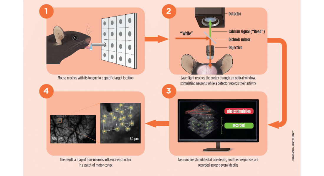
But technological advances in tools and methods have changed the game. These include two-photon excitation microscopy, which enables imaging inside the brains of living organisms, and optogenetics, which uses genetic modifications to make neurons fire in response to laser stimulation. By photostimulating neurons and imaging their responses, the connectivity between neurons in the living brain can be mapped without the need to slice a brain sample.

These tools pave the way for scientists like Arseny Finkelstein to map connectivity in a living brain over multiple time periods to study connectivity during memory formation, which Finkelstein is beginning to do now.

The method behind these studies, known as an all-optical approach for imaging and stimulating, uses genetic engineering in neurons to make them fire when struck by a minuscule amount of light delivered by laser, and to fluoresce when they fire.

Finkelstein’s method employs these recently established techniques to map the constellation of neurons in a mouse’s cortex, only he was able to dramatically speed up the process. The key advance was a new way to stimulate and image neurons at the same time. The researchers delivered long, randomly ordered flashes of light to activate many neurons while recording three-dimensional calcium signals that reveal which neurons are active. This let them map connections across a much larger network than before.

In practical terms, they could stimulate far more potential source neurons and monitor far more responding neurons at once. As a result, they were able to test a much larger share of the possible connections in the network than earlier methods allowed, and much faster — half a million neuron pairs in a mere 30 minutes, or 10 times the speed of previous mouse studies.

Instead of looking at the brain through a keyhole, Finkelstein could now glimpse it through a windowpane.


Subscribe to our newsletter

* indicates required

All fields are mandatory.